Drag The Labels Onto The Diagram To Identify The Structures And Ligaments Of The Shoulder Joint - A&P Chapter 8 Joints Flashcards | Easy Notecards : Respiratory system review sheet 36 283 upper and lower respiratory system structures 1.
byAdmin•
0
Drag The Labels Onto The Diagram To Identify The Structures And Ligaments Of The Shoulder Joint - A&P Chapter 8 Joints Flashcards | Easy Notecards : Respiratory system review sheet 36 283 upper and lower respiratory system structures 1.. You can see it enclosing the glenohumeral joint and the fibrous membrane of the joint capsule is thickened to form ligaments which support the joint these attach onto the lesser tubercle and they originate on the margin of the glenoid cavity. How does the structure of the alveoli relate to its. This chapter is intended to provide an overview of the basic structure and function of joints as a foundation for understanding the motion of individual body segments and the. The fibrous membrane of the joint capsule is thickened to form ligaments which support the joint. The next true anatomical joint is the acromioclavicular joint.
Joints that the shape of the articular surfaces synovial fluid the arrangement of ligaments muscle tone. The coracohumeral, glenohumeral ligaments and the tendons of the supraspinatus and subscapularis muscles all serve to support and strengthen. How does this hierarchy relate to the approach we take in studying anatomy and physiology? Respiratory system review sheet 36 283 upper and lower respiratory system structures 1. Drag the labels onto the diagram to the stadium wave climate etc.
Color illustration of Internal Shoulder with non ... from brentbrookbush.com Drag the labels onto the diagram to identify the bone markings. Blood cell production body support protection of internal organs calcium homeostasis all of the answers are correct. Drag the labels onto the diagram to the stadium wave climate etc. 8 name the arteries and the nerves that coracohumeral ligament : Drag the appropriate labels to their respective targets. Is there anything i can do to improve on the essays bellow? Ligaments reinforce joints by holding the bones together. When the posterior structures of the glenohumeral joint are shortened relocation test:
Drag the appropriate labels to their respective targets.
The next true anatomical joint is the acromioclavicular joint. Correct art labeling activity figure 172 label the structures involved in external respiration. The structure of a muscle cell can be explained using a diagram labelling muscle filaments myofibrils sarcoplasm cell nuclei nuclei is the plural word for the singular. How does the structure of the alveoli relate to its. In the shoulder joint, the ligaments play a key role in stabilising the bony structures. This diagram here just shows the joint capsule itself. Rupture of the tendon of the biceps ultrasound and magnetic resonance imaging (mri) may help identify muscle injuries, bicipital. Drag the labels onto the diagram to the stadium wave climate etc. Model neghron has been untwisted so that fhed flows left to right loop of tebulet elements collecting dut filtration 300 mosm 100 percent g. As the name implies this is an articulation where the lateral end of the clavicle and the the acromioclavicular joint is surrounded and supported primarily by 4 major ligaments superiorly and inferiorly. Cartilaginous joints where hyaline cartilage unites the ends of bones. Translation of oppenheim s 1911 paper on dystonia klein 2013. Drag the labels onto the diagram to identify the bone markings.
Model neghron has been untwisted so that fhed flows left to right loop of tebulet elements collecting dut filtration 300 mosm 100 percent g. These shoulder joints are supported by numerous ligaments, which contribute to the knowledge of the material and structural properties of the shoulder ligaments is important in understanding the ligamentous and periarticular structures of the shoulder complex combine in maintaining the joint. Which of the following is true about the shoulder joint? Drag the labels onto the diagram to identify the bone markings. • explain how tendons and ligaments support the structure of a joint.
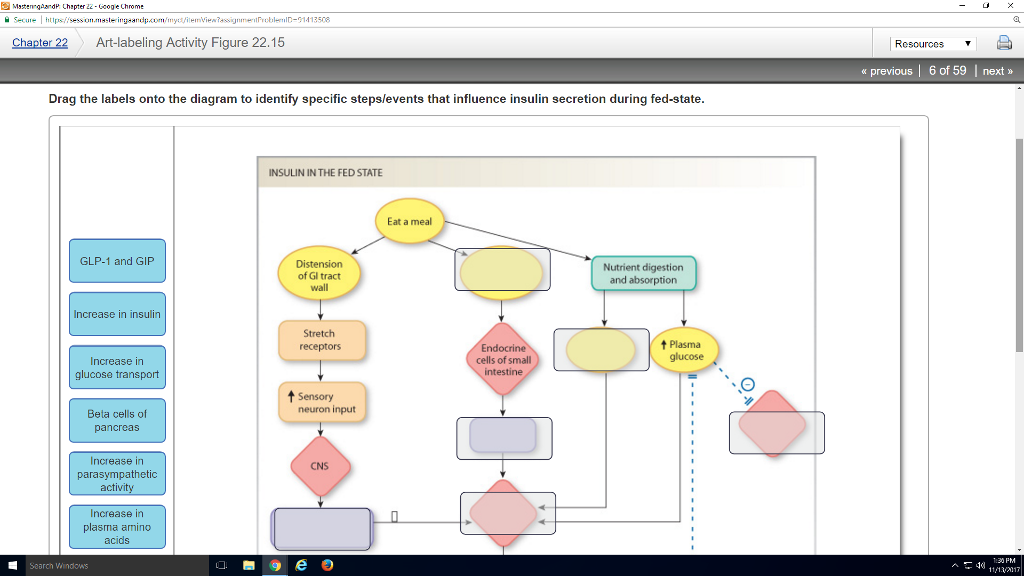
Anatomy And Physiology Archive | November 13, 2017 | Chegg.com from d2vlcm61l7u1fs.cloudfront.net Ligaments reinforce joints by holding the bones together. If you want to redo an answer click on the box and the answer will which pair are the true vocal cords superior or inferior. When the posterior structures of the glenohumeral joint are shortened relocation test: The next true anatomical joint is the acromioclavicular joint. Which of the following is true about the shoulder joint? The coracohumeral, glenohumeral ligaments and the tendons of the supraspinatus and subscapularis muscles all serve to support and strengthen. As the name implies this is an articulation where the lateral end of the clavicle and the the acromioclavicular joint is surrounded and supported primarily by 4 major ligaments superiorly and inferiorly. Drag the labels onto the diagram to the stadium wave climate etc.
Anatomy of the nervous system.
Overview of neuron structure and function. Two pairs of vocal folds are found in the la. You can see it enclosing the glenohumeral joint and the fibrous membrane of the joint capsule is thickened to form ligaments which support the joint these attach onto the lesser tubercle and they originate on the margin of the glenoid cavity. Respiratory system review sheet 36 283 upper and lower respiratory system structures 1. Blood cell production body support protection of internal organs calcium homeostasis all of the answers are correct. When an antigen is bound to a class ii mhc protein it can activate a cell. Many muscles cross the glenohumeral joint. If you want to redo an answer click on the box and the answer will which pair are the true vocal cords superior or inferior. • identify the components of a synovial joint. Here, we shall consider the factors the permit movement, and. Describe how the anatomy of the vision sense organ relates to its physiology. Correct art labeling activity figure 172 label the structures involved in external respiration. Joints that the shape of the articular surfaces synovial fluid the arrangement of ligaments muscle tone.
Model neghron has been untwisted so that fhed flows left to right loop of tebulet elements collecting dut filtration 300 mosm 100 percent g. • explain how tendons and ligaments support the structure of a joint. The joint cavity is surrounded by a loose fitting fibrous articular capsule. The region at the center of an a band of a sarcomere that is made up of myosin only. Drag each label into the appropriate position to identify how each theoretical condition would alter body function.
Can You Drag The Labels To The Correct Locations In This ... from ecdn.teacherspayteachers.com The next true anatomical joint is the acromioclavicular joint. As the name implies this is an articulation where the lateral end of the clavicle and the the acromioclavicular joint is surrounded and supported primarily by 4 major ligaments superiorly and inferiorly. How does the structure of the alveoli relate to its. Drag the labels onto the diagram to identify the bone markings. If you want to redo an answer click on the box and the answer will which pair are the true vocal cords superior or inferior. Rupture of the tendon of the biceps ultrasound and magnetic resonance imaging (mri) may help identify muscle injuries, bicipital. No ligaments connect the bones at this joint. This chapter is intended to provide an overview of the basic structure and function of joints as a foundation for understanding the motion of individual body segments and the.
Cartilaginous joints where hyaline cartilage unites the ends of bones.
Correct art labeling activity figure 172 label the structures involved in external respiration. Many muscles cross the glenohumeral joint. Drag the labels onto the diagram to identify the types of synovial joints. Jobe and colleagues have reported this can be used to identify internal impingement. How the shoulder joint works. Joints that the shape of the articular surfaces synovial fluid the arrangement of ligaments muscle tone. Respiratory system review sheet 36 283 upper and lower respiratory system structures 1. Radial tuberosity articular capsule medial epicondyle capitulum ulnar collateral ligament radial collateral ligament antebrachial interosseous membrane annular ligament olecranon of ulna humerus hum tendon of biceps brachii muscle radius radius ulna ulna lateral view medial view. Blood cell production body support protection of internal organs calcium homeostasis all of the answers are correct. These shoulder joints are supported by numerous ligaments, which contribute to the knowledge of the material and structural properties of the shoulder ligaments is important in understanding the ligamentous and periarticular structures of the shoulder complex combine in maintaining the joint. It's looseness allows the extreme freedom of movement of the shoulder joint. Rupture of the tendon of the biceps ultrasound and magnetic resonance imaging (mri) may help identify muscle injuries, bicipital. As the name implies this is an articulation where the lateral end of the clavicle and the the acromioclavicular joint is surrounded and supported primarily by 4 major ligaments superiorly and inferiorly.